[Updated] HKUMed reveals the electron microscope image of SARS-CoV-2 Omicron variant
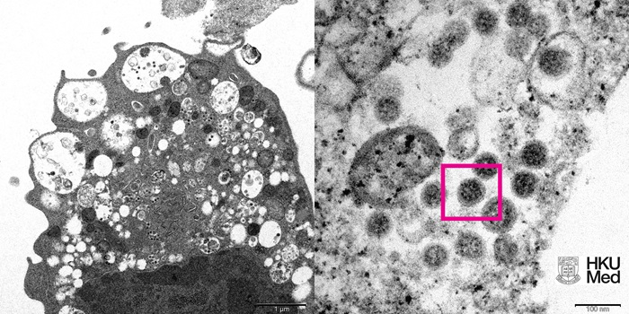
(Left) Low magnification electron micrograph of a monkey kidney cell (Vero E6) after infection with the SARS-CoV-2 Omicron variant showing cell damage with swollen vesicles containing small black viral particles.
(Right) High magnification electron micrograph of an infected Vero E6 cell showing aggregates of viral particles with corona shaped spikes on their surface (red box).
Photo credit: Professor John Nicholls, Clinical Professor of Department of Pathology; and Professor Malik Peiris, Tam Wah-Ching Professor in Medical Science and Chair Professor of Virology, School of Public Health, HKUMed; and Electron Microscope Unit, HKU.
High-resolution version of the image is available at: https://bit.ly/3GibEin
The appended information is updated on December 13, 2021
![]()
(Left) Low magnification electron micrograph of a Vero (monkey kidney) cell 24 hours after infection with the Omicron variant of SARS-CoV-2. The boxes show clusters of viral particles in the cytoplasm.
(Centre) High magnification electron micrograph of the larger box in the left panel showing aggregates of viral particles in vesicles and on the surface of the cell.
(Right) High magnification electron micrograph of the smaller box in the left panel showing large aggregates of viral particles with spikes (box) in membrane bound vesicles.
Photo credit: Professor John Nicholls, Clinical Professor of Department of Pathology, HKUMed; Professors Malik Peiris, Tam Wah-Ching Professor in Medical Science and Chair Professor of Virology, School of Public Health, HKUMed; Professor Leo Poon Lit-man, Professor and Head of Division of Public Health Laboratory Sciences, School of Public Health, HKUMed; and Electron Microscope Unit, HKU.
High-resolution version of the image is available at: https://bit.ly/3EPkKTx


.png)
